What is Golgi Neuron Silver Stain?
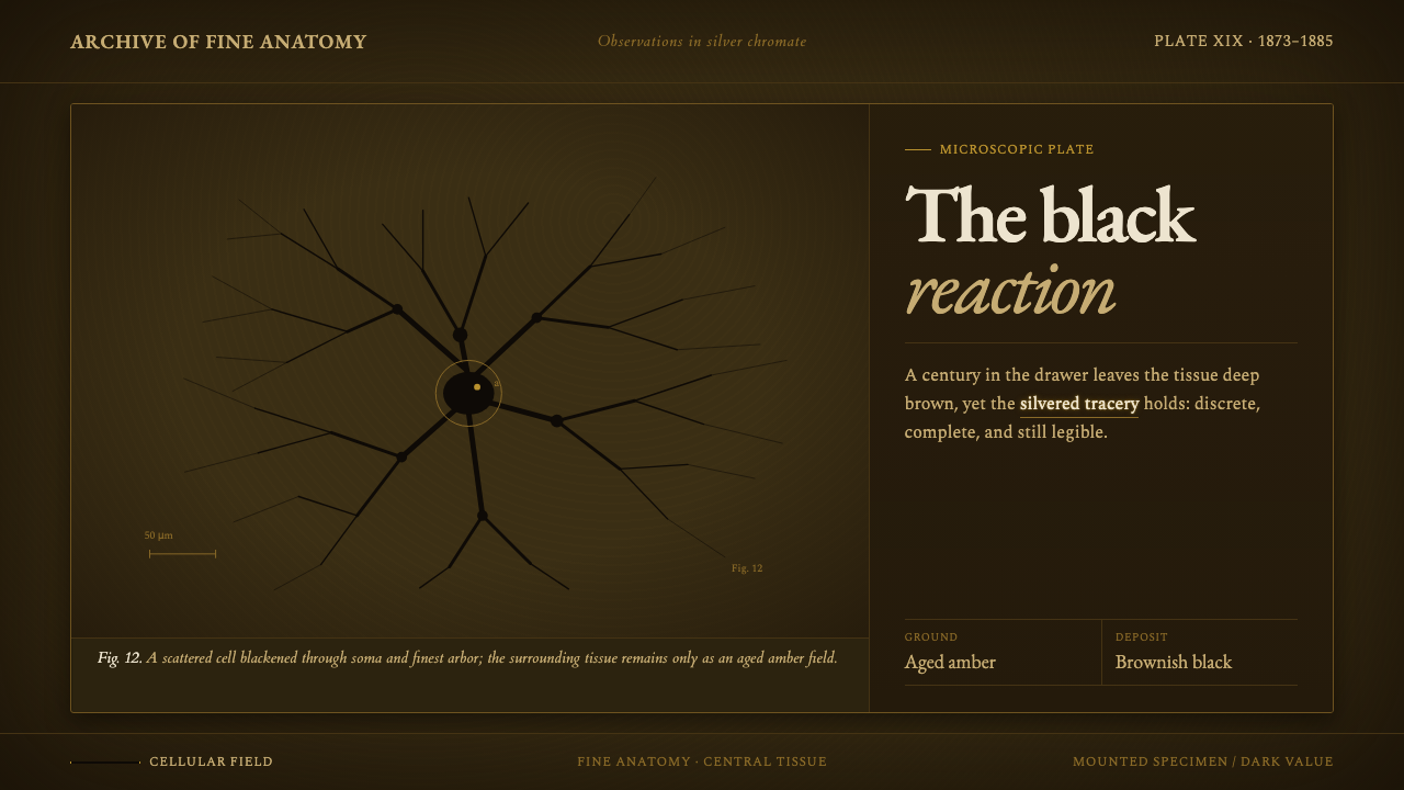
Golgi Neuron Silver Stain turns a nineteenth-century microscopic method into a dark archival language: near-black neural tracery spreading through an amber field that feels transparent, aged, and kept too long in a drawer.
Golgi Neuron Silver Stain in brief
Golgi Neuron Silver Stain is a visual system based on Camillo Golgi’s reazione nera, the black reaction developed in 1873. Silver chromate entered a scattered few neurons and darkened each soma, axon, and finest dendrite, leaving the neural form legible against transparent yellow tissue. This style reads that specimen after time has acted on it: the yellow has deepened into amber and brown, while the branching cells have settled into near-black tracery.
The central effect is not the spectacle of a bright scientific plate but the quiet authority of an old specimen. The field is dark, warm, and translucent rather than flatly black. Neural branches do not form a decorative pattern; they emerge as evidence, irregularly distributed and structurally specific. Empty space matters because the stain never fills the whole tissue. What appears is a partial revelation: a few cells made visible inside a larger, quieter ground.
The aesthetic depends on a disciplined tension between biological complexity and visual restraint. Dendrites may branch delicately, axons may travel with a different directional insistence, and somas may act as dense anchors, but every mark remains subordinate to the specimen logic. The result feels archival, neuroanatomical, and materially aged without becoming nostalgic illustration. It is a dark system whose richness comes from branching, transparency, and accumulated time.

Where does Golgi Neuron Silver Stain come from?
The method begins in Pavia, Italy, in 1873, when Camillo Golgi developed the reazione nera. Its defining visual event was selective darkness: silver chromate entered a scattered few neurons and blackened their complete visible structure, from soma to axon to the finest dendrite. The surrounding tissue remained transparent yellow, creating the contrast that made the method so distinctive. The visual language therefore begins not with a palette invented for decoration, but with a material relationship between stained cells and translucent tissue.
The historical setting is nineteenth-century histology, where the microscope made tissue legible through processes of preparation and contrast. Golgi’s method belonged to that scientific effort, but its image-making consequences are unusually strong. The stain did not turn the whole field into an even diagram. It allowed isolated neural structures to appear with exceptional completeness while the rest of the tissue receded. The resulting composition contains both precision and absence: a fully darkened cell suspended inside a larger field that remains only partly disclosed.
The method also belongs to the history of neuroanatomy. In 1885, it was associated with a monograph on the fine anatomy of the central nervous system, placing the black reaction within a sustained attempt to understand neural structure at microscopic scale. The visual system inherits that concentration. It treats each branch as anatomy rather than ornament and each empty interval as part of the evidence. A convincing application should feel studied, sectional, and patient, as though the image has been examined rather than merely styled.
The final reference point is Santiago Ramón y Cajal’s description of the image as “brownish black upon a transparent yellow background.” Golgi Neuron Silver Stain interprets that phrase through the value of a real specimen aged for a century: the transparent yellow becomes deep amber and brown, while the brownish black settles toward near-black. This is why the style feels darker than a fresh plate and warmer than a black-and-white diagram. It is a historical method translated into the visual memory of an old slide.
What defines the Golgi Neuron Silver Stain look?
Amber Ground
The ground begins as transparent yellow tissue but is read through age: warm amber, deep brown, and the suggestion of translucency. It should feel like material seen through a microscope slide, not like a flat decorative background. The field carries most of the atmosphere, so its darkness must remain open enough for the neural tracery to appear inside it rather than sit on top of it.
Near-Black Tracery
Neurons are rendered in a brownish near-black rather than a sterile absolute black. Somas form dense visual anchors; axons extend with directional force; dendrites divide into increasingly fine branches. The marks should preserve the feeling of a complete stained structure, with variation in density and branching instead of a repeated ornamental motif.
Selective Revelation
Only a scattered few neural forms should command attention. The method’s character comes from what becomes visible and what remains unresolved. A field crowded with equal branches loses the logic of the specimen. Strong compositions allow one cell or a small cluster to emerge while the surrounding amber tissue remains quiet, transparent, and only partially described.
Branching Direction
The visual rhythm is organic but not soft. Dendrites spread, split, and taper; axons suggest travel across the field; somas interrupt the movement with compact weight. Direction should feel discovered inside the tissue rather than imposed by a decorative pattern. Branching is the primary source of motion, replacing gradients, ornament, or theatrical lighting.
Aged Contrast
Contrast is warm and archival rather than stark and digital. Near-black cells must remain legible against deep amber, but the relationship should never become a simple black-on-white diagram. The field and the stain belong to the same aged material world, separated by density and value while remaining related in temperature.
Specimen Honesty
Every visual choice should suggest a slide, a stain, and tissue under observation. The style has no need for polished metallic effects, ornamental frames, or artificial depth. Its authority comes from accepting the evidence of the specimen: transparent ground, darkened cells, incomplete revelation, and the quiet irregularity of biological branching.

Who shaped Golgi Neuron Silver Stain?
Camillo Golgi developed the reazione nera in Pavia in 1873. Within this visual language, he represents the act of making hidden neural structure visible through selective staining. The dark soma, axon, and dendrite are not separate graphic inventions; they are the visible consequence of the method associated with his name.
Santiago Ramón y Cajal provides the defining verbal image for the system: “brownish black upon a transparent yellow background.” His phrase clarifies the essential relationship between the stained neural tracery and the tissue around it. The style follows that relationship into an older, darker register without abandoning the transparent yellow ground.
The reazione nera, or black reaction, is the method figure at the center of the style. Silver chromate enters a scattered few neurons and darkens their visible structure against transparent yellow tissue. As a design source, it supplies the system’s selective contrast, biological branching, and refusal of uniform coverage.
How do you use Golgi Neuron Silver Stain today?
For presentation slides, use the style as a complete specimen sequence. The cover should establish the amber field and introduce one dominant near-black neural structure, allowing the title to sit with the authority of a catalogued slide. Content slides should preserve the same tissue logic: quiet grounds, restrained type, and branch-like lines used only when they clarify structure. Data slides can treat each chart as a specimen field, with one dark mass or path carrying the main finding while secondary information recedes into the amber atmosphere.
For web interfaces, Golgi Neuron Silver Stain suits research dashboards, microscopy archives, neuroanatomical references, and analytical tools that need depth without theatrical gloss. Panels should feel like windows into a larger tissue field rather than isolated bright cards. On pricing pages, the same logic can distinguish tiers through density and clarity: the primary offer becomes the most legible stained structure, while supporting details remain quieter. Navigation should stay calm and typographic, with branching used sparingly as a locator or state indicator.
For editorial and marketing work, the style supports stories about science, archives, medicine, observation, and the persistence of knowledge. A feature article can open with a large dark neural form crossing an amber field, then shift into narrower reading areas where small diagrams and pull quotes behave like isolated cells. Marketing pages can use the contrast between a near-black specimen and aged tissue to make a product feel exacting and research-led. The strongest campaigns let the material atmosphere carry the emotion instead of adding sentimental imagery.
The common mistake is to turn the style into generic dark mode with amber accents. A black background, glowing orange lines, and abundant decorative branches do not recreate a Golgi specimen. The method depends on selective revelation, transparent tissue, and the uneven completeness of biological staining. Another mistake is making every neuron equally prominent. The field should contain hierarchy: a few structures become evidence, while the surrounding darkness remains an active, partially hidden ground.

Golgi Neuron Silver Stain — FAQ
Is Golgi Neuron Silver Stain meant to look like a bright scientific diagram?
No. Its reference is a specimen aged in darkness. The original relationship is brownish black on transparent yellow, but this system deepens the yellow into amber and brown and lets the neural forms settle toward near-black. It should feel like a real slide remembered after long storage, not a clean modern plate or a luminous medical interface.
Why is the background amber instead of simply black?
Because the tissue is part of the method’s identity. The black reaction darkened selected neurons against transparent yellow tissue. Removing that tissue relationship would reduce the style to dark decoration. Amber preserves the sense of a translucent specimen and gives the near-black branches a related but clearly stronger density.
Should every branch be visible and equally detailed?
No. Selective revelation is essential. The source method made a scattered few neurons visible while the surrounding tissue remained less resolved. A convincing composition therefore gives priority to one cell or a small cluster, while other traces may recede into the amber field. Equal visibility would create a pattern rather than a specimen.
Can the style be used for products unrelated to neuroscience?
Yes, if the product benefits from an atmosphere of observation, evidence, archival depth, or intricate structure. It can support research tools, knowledge products, archives, and analytical experiences. The application should preserve the method’s underlying behavior: a quiet material field, selective emphasis, and complexity that emerges through branching rather than decoration. Without those principles, only the amber-and-black surface remains.
Related design styles
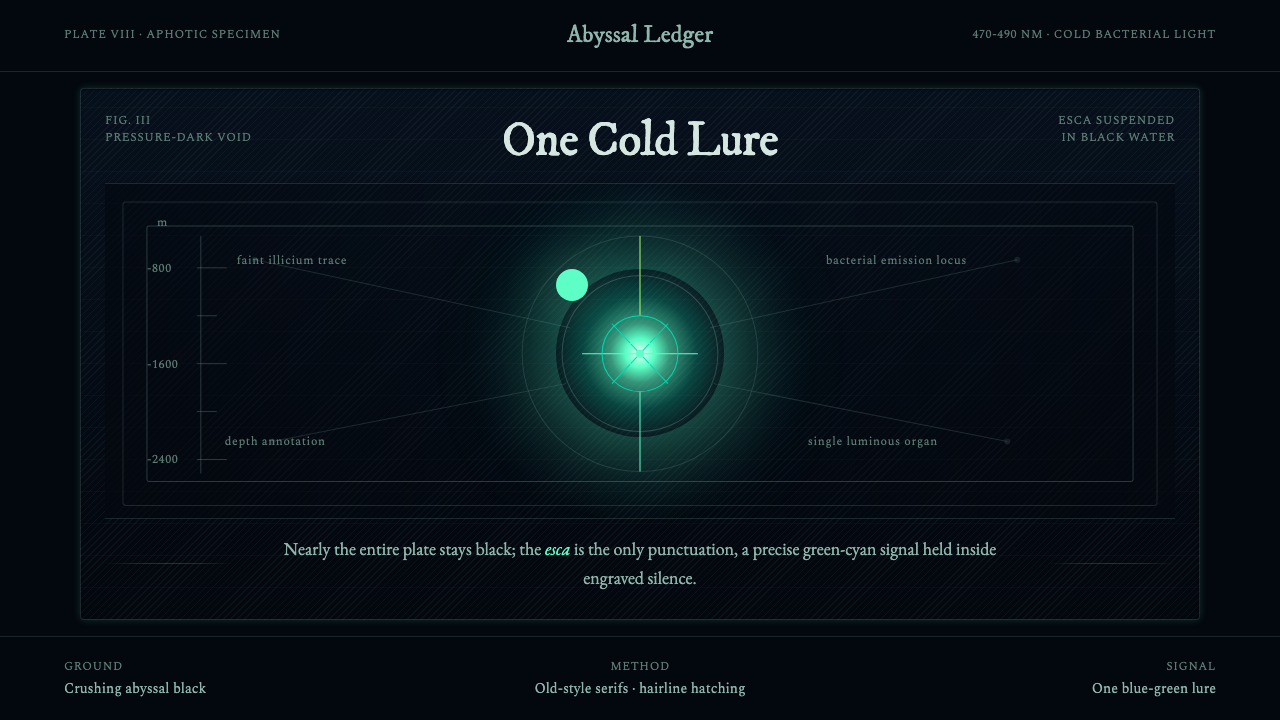 Anglerfish Lure BioluminescenceLight is scarce. Old serifs and hairline rules frame one cold green-cyan lure.
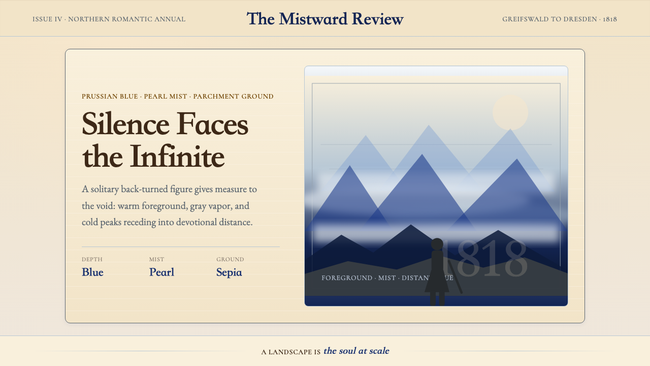 Caspar David FriedrichSilence becomes vast. Prussian blue depth, pearl mist, and parchment framing…
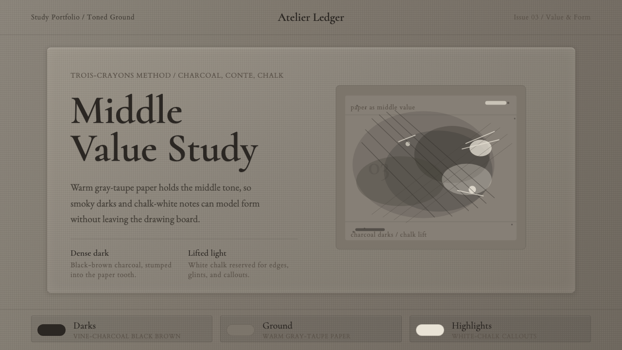 Charcoal & Conté DrawingAtelier calm, in middle value. Gray-taupe tooth, smoky charcoal, and chalk-wh…
 Claude Monet ImpressionistLight becomes the subject. Buttercream, pastel dapples, and Cormorant serif s…
 Degas Ballet PastelsBackstage glamour is labor. Jade walls, rose footlights, cropped serifs on cr…
 Gotland Picture StoneMeaning emerges slowly. Weathered limestone greys divide a story into shallow…